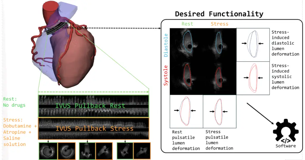
Coronary artery anomalies (CAA) with an intramural course are associated with elevated risks of ischemia and sudden cardiac death under stress. Intravascular ultrasound (IVUS) is essential for assessing coronary vessel dynamics in these patients. However, the rarity of such anomalies, along with unique geometric changes in the intramural course and ostium, complicates image analysis, leading to inconsistencies and time-consuming evaluations. Our developed executable, zero/low-code software addresses these limitations by providing automated lumen segmentation and cardiac phase identification in IVUS images acquired during rest and stress protocols.
The software includes: (1) Automated segmentation of lumen contours trained on 9,418 frames (developed by using human in the loop active learning process) validated on 691 frames and tested on 632 frames, IVUS frames from 76 patients (152 studies) with right CAA using a deep learning (DL) model; (2) Extraction of systolic and diastolic frames via a dual-gating approach combining image- and contour-based methods; and (3) A graphical user interface enabling manual correction of the results. The gating module was validated using a custom flow-loop simulating patient-specific hemodynamics, while segmentation accuracy was assessed via intraclass correlation coefficient (ICC) analysis comparing AI-generated contours with those delineated by experienced readers.
The DL model achieved a mean Dice score of 0.91 (SD: 0.08), sensitivity of 0.95 (SD: 0.12), and specificity of 1.00 (SD: 0.00) on the test set. ICC values for lumen area measurements were 1.00 (95%CI: 1.00–1.00) for rest and 1.00 (95%CI: 1.00–1.00) for stress conditions. The gating module demonstrated excellent reproducibility for identifying systolic and diastolic frames under both conditions (ICC = 1.00 for all).
AIVUS-CAA offers a reliable, automated tool for precise IVUS analysis at rest and during stress, enhancing the evaluation of geometrical changes of coronary vessels in CAA patients and enabling efficient clinical analysis in a streamlined workflow.