Summary: High-performance toolkit for aligning and fusing cardiac imaging from CCTA, IVUS, OCT, and MRI
Building a reliable three-dimensional model of a coronary artery requires combining imaging methods that are complementary but incomplete on their own. Intravascular imaging, acquired by a catheter inside the vessel using either ultrasound or light, offers exceptional local resolution but gives no whole-vessel context and no three-dimensional orientation. Coronary computed tomography angiography, a non-invasive scan, provides the global geometry but has limited spatial resolution and is affected by artifacts such as blooming. multimodars registers and fuses the two into one geometry. Its most demanding computational steps are written in Rust, a compiled programming language, which makes registration considerably faster than a pure Python implementation while the toolkit itself remains usable from Python.
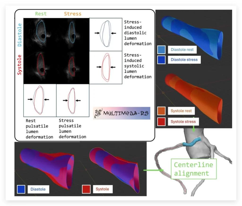
Intravascular geometries are registered using the Hausdorff distance, a measure of how far apart two shapes are, applied to vessel contours and catheter centroids, with four modes available: full, double-pair, single-pair and single. The result is then aligned onto a centerline derived from the computed tomography scan, either through a three-point anatomical scheme using the aortic, cranial and caudal directions, or by manual rotation. Centerlines are resampled to the contour spacing, centroids are translated to matched points and surface normals are aligned. The computed tomography geometry is automatically labeled by vessel region and morphed along the centerline to match the intravascular measurements, after which the two are stitched together, replacing a section of the lower-resolution model with the high-resolution one.
The toolkit was originally motivated by the need to quantify dynamic deformation of the vessel lumen in coronary artery anomalies, where comparisons at rest and under stress are diagnostically important. In a published case of an anomalous aortic origin of a coronary artery, the fused reconstruction revealed a compression pattern that was not visible on either imaging method alone. Beyond that setting, the single-pair mode supports general coronary artery disease workflows such as comparing the lumen before and after stent implantation, and the single mode reconstructs standalone geometry from any clinical pullback. High-fidelity lumen models of this kind are also a prerequisite for patient-specific blood flow and fluid-structure interaction simulations, and for digital twin and three-dimensional printing workflows used in device testing.
The package installs from the Python Package Index and accepts comma-separated value files, including those produced by the lab’s AIVUS-CAA software, as well as raw arrays, so that any external segmentation tool can be connected through its bidirectional interface. Documentation, example notebooks and an application programming interface reference are published online, together with utilities for contour smoothing, metric calculation and geometry export for visualization. multimodars is developed and maintained by the AI-CVM lab team and was published in the Journal of Open Source Software in 2026.
Cite as
Stark et al., (2026). multimodars: A Rust-powered toolkit for multi-modality cardiac image fusion and registration. Journal of Open Source Software, 11(121), 10131, https://doi.org/10.21105/joss.10131